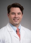
Day 2 :
Keynote Forum
Po-Kang Lin
National Yang Ming University, Taiwan
Keynote: A Universal liquid contact lens for vitrectomy
Time : 9.45-10.30

Biography:
Dr Po-Kang Lin has completed his MD from National Yang Ming University, Taiwan, and phD from Ins. of Bioelectornics and Bioinformatics, National Taiwan University, Taiwna. Currently, he is an associate professor of National Yang Ming University, and a clinical professor of National Defense Medical University, Taiwan. He also serves as an attending physician at the Department of Ophthalmology, Taipei Veterans General Hospital, Taiwan and as the director of ophthalmological ward. He is also a researcher at Biomedical Electronic Translation Research Center of National Chiao Tung University, Taiwan.
Abstract:
Usually we use different kinds of contact lens to perform vitrectomy. The contact lens changing during vitrectomy is tedious and labor consuming. To facilitate more efficient vitrectomy, we developed a contact lens universally substituting all kinds of contact lens for vitrectomy. The liquid lens could be put flat and served as a plano or concave contact lens. While tilted, it became a prism contact lens. With different angle of tilting, it can be used as a prism contact lens with a designated prism diopter. It could also be used under gas tamponade. While doing vitrectomy, the liquid contact lens could be applied all the way without changing. Universally, this unique liquid lens can replace plano, concave, 15-degree prism contact lens, 30-degree prism contact lens, and 45-degree prism contact lens for vitrectomy.
Keynote Forum
Sanghamitra Burman
Sight Years Eye Clinic, India
Keynote: Cataract Surgery In High Risk Corneas
Time : 9.45-10.30

Biography:
Dr. Sanghamitra Burman is a Consultant Eye Surgeon in Bangalore, India. She is a Fellowship trained specialist in Cornea, Cataract, Implant and Laser Refractive Surgery. Her areas of special interest include laser vision correction, keratoconus treatment, refractive lens surgery, keratoplasty, limbal stem cell transplantation, ocular surface disorders and reconstruction. She has extensive work experience at some of the world’s best eye institutes including Moorfield’s Eye Hospital, London, AIIMS, New Delhi and Aravind Eye Hospital, Sankara Nethralaya and LV Prasad Eye Institute. Dr Burman has publications in reputed journals and presented at various national and international conferences.
Abstract:
Keynote Forum
Melike Ozgun Gedar
Bahcesehir University School Of Medicine, Turkey
Keynote: Lower eyelid cadaveric anatomical dissection techniques

Biography:
Dr. Gedar is an Assistant Professor in Bahcesehir University Faculty of Medicine Department of Ophthalmology since 2014. She also have been giving clinical skills lectures to medical students in Bahcesehir University. Dr. Gedar is a currently a part-time consultant ophthalmic oculoplastic and orbital surgeon at the Dunya Goz Eye Hospital, one of the largest facilities of its kind in Istanbul. She is a specialist in all aspects of eyelid, lacrimal (tear drainage), eye socket and orbital disease.She has written a number of research papers relating in ophthalmology and has presented at both national and international meetings.
Abstract:
Lower Lid Blepharoplasty-Entropion-Ectropion Oculoplastic Surgery Cadaveric Dissection Course’ was performed in Bahcesehir University School of Medicine on February 19th, 2017 with the participation of 20 ophthalmologists. Lower eyelid anatomy, the lower eyelid entropion-ectropion surgical techniques, subciliary incision and transconjunctival approach to lower eyelid blepharoplasty, complication prevention and management methods are shown on the fresh frozen cadaveric eyelids. The surgical microscopic images linked to the master operation table have been recorded. The stages of the lower eyelid cadaveric dissection are presented together with details of the anatomical folds through video images. Cadaveric workshops as a primary modality of simulation based surgical skills training have been used for a few years in Turkey. Cadaveric dissection training in oculoplasty confers greatest benefit to the surgeons, despite disadvantages of tissue loss and form, degeneration of anatomical key points, diversification in the structure, lack of experience in live tissue tonus and bleeding. Also there are difficulties in providing cadavers and expense. It is believed that cadaveric dissection is the gold standard technique for surgical skill transfer in eyelid surgery.
- Ophthalmic Surgery | Ophthalmology | Refractive Eye Surgery | Types of ophthalmic Surgery | Ocular Oncology | Pediatric Ophthalmology | Intraocular Melanoma | Neuro-ophthalmology
Location: Yamato
Session Introduction
Tong Qiao
Shanghai Jiao Tong University, China
Title: Exploring The Causes And Secondary Procedure Chioce Of Consecutive Esotrooia After Surgery In Intermittent Exotrooia
Time : 11.30-12:00

Biography:
Abstract:
To describe the clinical features of congenital double elevator palsy (CDEP) and to evaluate different surgical outcomes based on improvements in the primary eye position and ocular motility.Sixteen patients with congenital double elevator palsy in Shanghai Children's Hospital were enrolled from July 2014 to January 2017. Forced duction test ( FDT ) was negative in 15 cases. Twelve patients underwent standard Knapp procedure, with or without horizontal squint procedure; one patient underwent Hummelsheim procedure (part of the tendons capsule transposed); two patients underwent augmented Knapp procedure.

Biography:
Dr. Christopher. B. Chambers earned his bachelor of science at the University of Notre Dame. He went to medical school at The Ohio State University before completing his residency at the Kresge Eye Institute where he served as chief resident. Following residency Dr. Chambers completed an American Society of Ophthalmic Plastic and Reconstructive Surgery (ASOPRS) fellowship at the University of Pennsylvania and the Children’s Hospital of Philadelphia. Dr. Chambers was the associate residency director while on staff at Northwestern University for four years. Dr. Chambers is the ASOPRS fellowship director and associate residency director at the University of Washington Department of Ophthalmology. Dr. Chambers has won medical student teaching awards and served as a team physician for the NHL Chicago Blackhawks. Dr. Chambers is active in national and local leadership and is the Chair of the YASOPRS committee as well a member of the ASOPRS education committee.
Abstract:
This talk will discuss expansion of the micropthalmic orbit to help in orbital and hemifacial growth. The talk will cover the relevant anatomy and techniques available to provide soft tissue and bone growth through dynamic stimulation of the tissue.
Biography:
Dr. Ravi Dhar Bhandari is an ophthalmologist at Geta Eye hospital in Nepal
Abstract:
Objective: To compare the outcome of dacryocystorhinostomy surgery with silicone tube intubation and without silicone tube intubation at Geta eye hospital.
Method: A hospital based retrospective comparative case study in which 87 subjects operated for dacryosystitis were analyzed, of which 49 were with silicone tube intubation and 38 were without silicone tube intubation. Study data were obtained from hospital records in which subjects were followed up on one week, one and half month and three months, postoperatively.
Result: On three months of surgery, 72 of 87 (82.76%) were followed up. Of 39 with silicone tube 35 (89.7%) had patent ducts and of 33 without silicone tube 29 (87.9%) had patent ducts on lacrimal syringing which was considered as success of the surgery. Only 4 patients in each group, 10.3% silicone tube and 12.1% without silicone tube had regurgitation of mucous or pus (failure) on lacrimal syringing. Both the group did not differ significantly (p=0.54).
Conclusion: Dacryocystorhinostomy with silicone tube intubation and without silicon tube intubation procedure offer similar efficient outcome.
